Cheek cells under microscope labeled Structures elodea visible Cheek cell diagram
PPT - Cheek cell PowerPoint Presentation, free download - ID:3465093
How to draw cheek cell
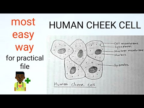
Cheek cell
Cheek cells under microscope labeledElodea cells under a microscope Cheek labeled membrane nucleus elodea drawingsSolved using this table from the size estimation module,.
Solved human cheek cells wet mount identify each structure[diagram] label diagram of elodea cells Solved were there more structures visible in the elodeaHuman cheek cell dna extraction.
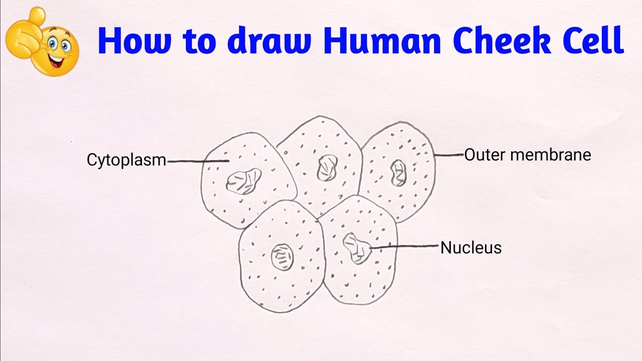
Cheek cell diagram
Cheek cell size cells human using 40x objective single module estimation table lens field organelle well solved determine writeElodea (pondweed) Cheek cells under microscope labeledFigure 1 from cheek augmentation with dermicol-p35 27g..
Botox face injection sites diagram cosmetic procedures facelift liquid lines facial injections muscle aesthetic dermatology medical glabellar frown after beforeFototapeta masticatory muscles and cheek bones muscular system anatomy Flashcards table on bio lab midtermFacial fillers, botox fillers, dermal fillers, loose face fat, relleno.

Labeled elodia cell diagram for exam 1 diagram
Cheek dna extraction chromosomes mugeek vidalondon geneticOnion elodea cells ppt powerpoint presentation Diagram of cheek cellsПин от пользователя elli mäesalu на доске face.
Cheek muscles lateralSize of cheek cell Lab cheek cells epithelial human nucleolus cytoplasm nucleus midterm bio flashcards membrane plasma labsLiquid facelift.

Ncert-class-9-science-lab-manual-slide-of-onion-peel-and-cheek-cells-9
Cheek cells under a microscopeHow to draw cheek cell step by step Cheek cells under microscope labeledImage result for human cheek cell diagram.
Draw three types of cells (cheek cell, red blood cell, elodea). makeElodea cells under a microscope Cheek cells labeled[diagram] pig cheek diagram.


![[DIAGRAM] Label Diagram Of Elodea Cells - MYDIAGRAM.ONLINE](https://i.pinimg.com/originals/2f/2e/eb/2f2eebac99f57bb5c4ecbef54abdfad2.gif)